Ask your patients
Have you had
outbreaks
of boils
during the
last 6 months, and if
so,
where and how
many?
If yes, it
could be HS
Up to ~90% sensitivity in
evaluation5
There are 3 essential diagnostic criteria that can
help distinguish HS from other skin conditions6
Lesions
One or more painful, inflammatory,
deep-seated nodules, abscesses, or
comedones.3
Location
Lesions are most common on flexural sites,
like the axillae, inframammary, groin, perineal
and perianal regions. They are less common
on ears, face, scalp, back, chest, or legs.3,6
Chronicity
Recurrence at least twice within 6
months and/or persistence of at
least 3 months2,7

Commonly Involved Areas3,6
-
Inframammary region
-
Groin
-
Axillae
-
Buttocks and perianal
region -
Perineal region
Less common Areas2 Around the ears, back, face, scalp, chest, and legs
Know HS when you see it
Early HS lesions may resemble other dermatological conditions. Knowing the common presentations, common differential diagnoses, and
stage of disease can help you identify HS more quickly and make referrals to HS-treating dermatology providers sooner.2
-
Abscess
Image of abscess of HS patient provided by
Dermatology Online Journal, copyright Noah Scheinfeld,
MD. -
Nodule
Image of nodule of HS patient provided by DermNet.
-
Follicular papule/pustule
Image of follicular papules/pustules of HS patient
provided by Dermatology Online Journal, copyright Noah
Scheinfeld, MD. -
Tunnel
Image of sinus tract/fistula of HS patient provided by
Experimental Dermatology. -
Scar/cord
Image of cord/scar of HS patient provided by
Dermatology Online Journal, copyright Noah Scheinfeld,
MD. -
Comedone
Image of comedones of HS patient provided by
American Family Physician.
-
Folliculitis
Image provided by International Journal of Dermatology.
-
Carbuncle
Image provided by Science Source.
-
Cutaneous Crohn's disease
Image provided by British Medical Journal.
-
Acne vulgaris
Image provided by Journal of Clinical and Aesthetic Dermatology.
-
Intergluteal pilonidal cysts
Image provided by Science Source.
-
Pyogenic granuloma
Image of granuloma provided by iStock.
-
Mild
Image of inframammary fold of HS patient provided by Dr Ali Moiin.
Sometimes described as Hurley Stage 1.
Typically presents as 1 or more nodules
with no to minimal scarring or tunnel
formation. -
Moderate
Image of axilla of HS patient provided by Science Source.
Sometimes described as Hurley Stage 2.
Typically presents as multiple nodules along
with limited tunneling and/or scarring. -
Severe
Image of axilla of HS patient provided by Science Source.
Sometimes described as Hurley Stage 3.
Typically presents as multiple or
extensive interconnected tunnels, nodules,
and scarring.
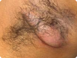
HS can appear differently depending on skin tone19
Hidradenitis suppurativa disproportionally affects patients of color and is associated with increased disease severity. Knowing the clinical
presentations of HS on darker skin tones may help you accurately identify HS.4,19
Presentations of HS unique to patients of color

Subtle erythema20
Image of axilla of HS patient provided by Science Source.

Violaceous, dark brown, and gray lesions20
Image of groin of HS patient provided by Dr Ali Moiin.

Post-inflammatory hyperpigmentation1,21
Image of axilla of HS patient provided by Science Source.

Keloids22
Image of chest provided by Science Source.
Hear from your peers about identifying and diagnosing HS

1665646103
1_kqu2whou
The healthcare providers featured in these videos were compensated for their time. These videos represent their opinions and do not represent the opinions of Novartis.

Help shine a light on HS
by sharing
on social
media today
You can find HS care near you
or your patients
using our care
locator
Discover strategies that can shed
light
on HS management and
treatment
References: 1. Sabat R et al. Nat Rev Dis Primers. 2020;6(1):18. 2. Saunte DML et al. JAMA. 2017;318(20):2019-2032. 3. Kimball AB et al, eds. Hidradenitis Suppurativa: A Disease Primer. Adis; 2017. 4. Kilgour JM et al. JAAD Int. 2021;3:42-52. 5. Vinding GR et al. Br J Dermatol. 2014;170(4):884-889. 6. Revuz J. J Eur Acad Dermatol Venereol. 2009;23(9):985-998. 7. Zouboulis CC et al. Exp Dermatol. 2021;30(suppl 1):27-30. 8. Fitzpatrick TB. Arch Dermatol. 1988;124(6):869-871. 9. Scheinfeld N. Dermatol Online J. 2014;20(4):22373. 10. Daxhelet M et al. Dermatology. 2020;236(5):431-438. 11. Martorell A et al. Dermatol Surg. 2019;45(1):1237-1244. 12. Frew J et al. JAMA Dermatol. 2021;157(4):449-455. 13. Elkin K et al. Skin Res Technol. 2021;26(1):11-19. 14. Margesson LJ et al. Best Pract Res Clin Obstet Gynaecol. 2014;28(7):1013-1027. 15. Micheletti RG. Semin Cutan Med Surg. 2014;33(suppl 3):S48-S50. 16. Winters RD, Mitchell M. Folliculitis. In: StatPearls. Treasure Island (FL): StatPearls Publishing; 2022. PMID: 31613534. 17. Deckers IE et al. Curr Derm Rep. 2014;3(1):54-60. 18. Alikhan A et al. J Am Acad Dermatol. 2019;81(1):76-90. 19. Okeke CAV et al. Dermatology. 2022;238(1):180-184. 20. Heath CR et al. Cutis. 2022;110(3):159-160. 21.Taylor S et al. J Cutan Med Surg. 2009;13(4):183-191. 22. Jfri A et al. JAAD Case Rep. 2019;5(8):675-678.
4/23 262711